
Top 3 Alternatives of Sectra for Advanced 3D Tooling
Many teams still rely on Sectra for 3D imaging but now face delays when specialists need remote access or when studies must move between sites.
These access gaps slow surgical planning and force extra steps for sharing large files. This article lays out three concrete criteria to judge any 3D tool against your workflow, explains how Medicai meets those criteria, and ranks the top two remaining options so you can decide which upgrade best fits your current setup.
What to Look For in Advanced 3D Tooling Solutions
Advanced 3D tooling must deliver accurate multiplanar reconstruction, measurement precision, and regulatory compliance without forcing radiologists and surgeons to master multiple disconnected systems.
Procurement teams evaluate six minimum technical requirements when selecting medical imaging platforms.
Sub-millimeter measurement accuracy on CT/MRI forms the foundation of reliable diagnostic work. Teams should ask vendors for documented precision testing on phantom studies and real patient datasets.
Real-time volume rendering at 60 fps enables smooth navigation through complex anatomical structures. Questions about GPU requirements and frame rate consistency across different workstation configurations help verify this capability.
Native DICOM/HL7 interoperability ensures seamless data exchange with existing radiology software and PACS alternatives. Procurement teams must confirm support for standard DICOM compliance and HL7 integration protocols.
HIPAA/GDPR encryption at rest and in transit protects patient data throughout the imaging workflow. Direct questions about encryption standards, key management, and audit trail capabilities reveal security implementation details.
FDA/CE clearance documentation validates regulatory compliance for clinical use. Teams should request current clearance certificates and review any limitations on approved indications.
Zero-footprint browser access eliminates installation requirements and simplifies deployment across hospital networks. Questions about supported browsers and performance on standard hardware configurations clarify implementation needs.
1. Medicai - Best Overall
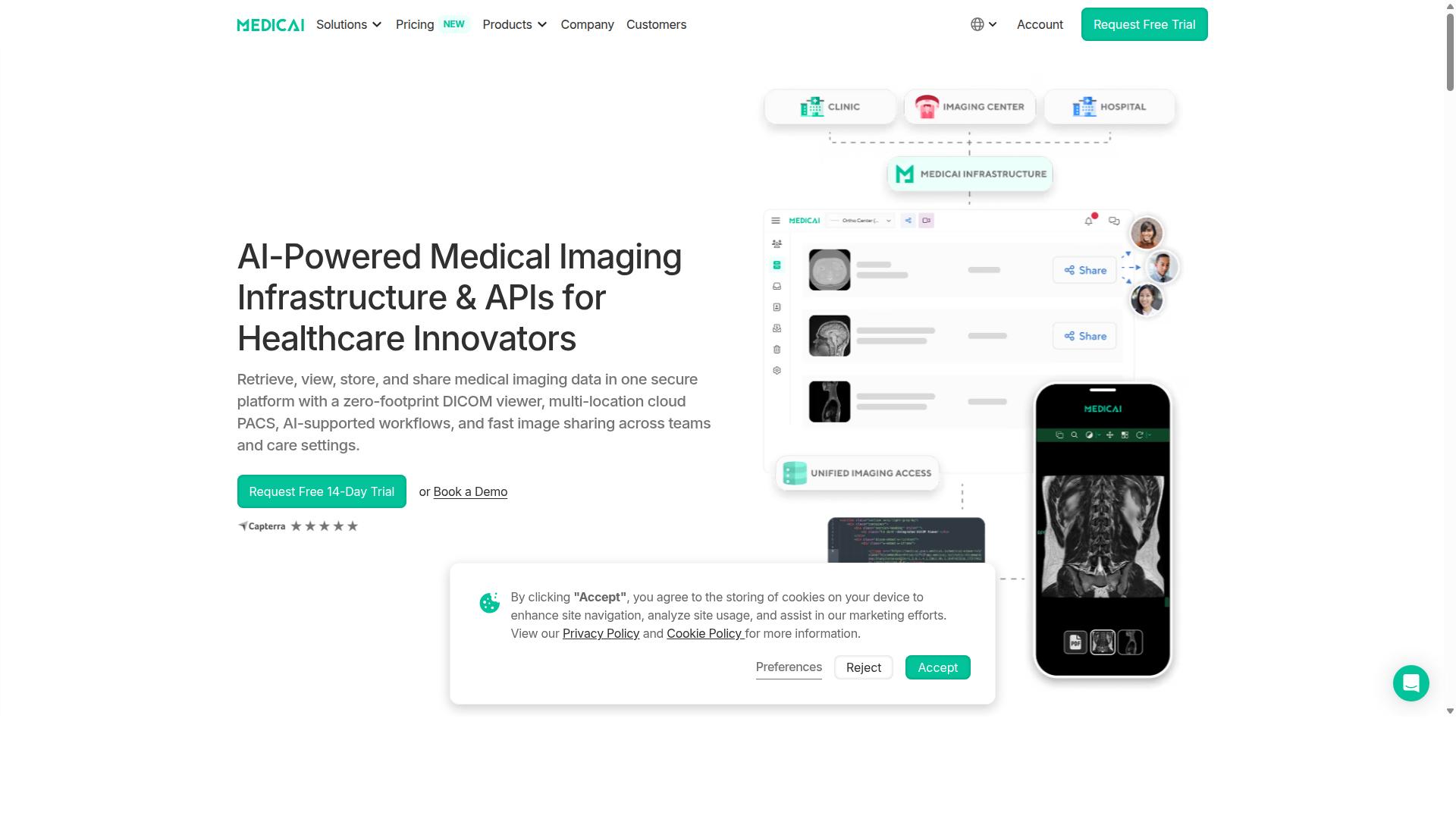
Medicai combines a zero-footprint DICOM viewer with cloud PACS, vendor-neutral archive, and global image exchange, positioning it as a platform that handles 3D reconstruction, surgical planning, and multi-location collaboration from a single HIPAA-compliant workspace.
The platform runs entirely in the browser without requiring on-premise servers. Teams access the same datasets from different locations while maintaining full 3D tooling performance.
Integration with existing hospital systems occurs through standard interoperability standards. This architecture removes hardware maintenance costs while preserving diagnostic quality across all workflows.
Cloud PACS with Advanced 3D Visualization
The platform renders CT and MRI datasets directly in the browser using server-side GPU acceleration, eliminating the need for local workstations to download full studies.
Users activate 3D volume rendering by selecting a study from the cloud PACS interface. They switch to the 3D tab to access volume rendering and multiplanar reconstruction tools without installing any plugins.
Window presets adjust automatically based on tissue type, while STL file export supports surgical navigation systems. The zero-footprint approach means any approved device can handle these tasks through standard web browsers.
AI-Supported 3D Workflows and Structured Reporting
AI models automatically segment anatomical structures, calculate bone density, and pre-populate implant templates so surgeons spend minutes instead of hours on preoperative planning.
The workflow begins with CT upload to the cloud PACS. AI segmentation completes within seconds, generating editable 3D models that users refine by adjusting anatomical landmarks.
Orthopedic templating integrates with major implant manufacturers through the structured reporting system. Measurements update automatically as users modify the 3D models, maintaining accuracy throughout the preoperative planning process.
Multi-Location Image Sharing for Surgical Planning
A single secure link gives referring surgeons, radiologists, and device reps simultaneous access to the same 3D dataset with synchronized cursors and annotations.
Users generate case links through the platform interface and set 30-day expiration periods. Role-based permissions control whether recipients can view, annotate, or download the shared content.
API endpoints push HL7 messages directly to hospital EMRs, removing manual upload steps. This process supports the 70 clinics and hospitals currently using the platform for collaborative surgical planning across different locations.
2. PostDICOM
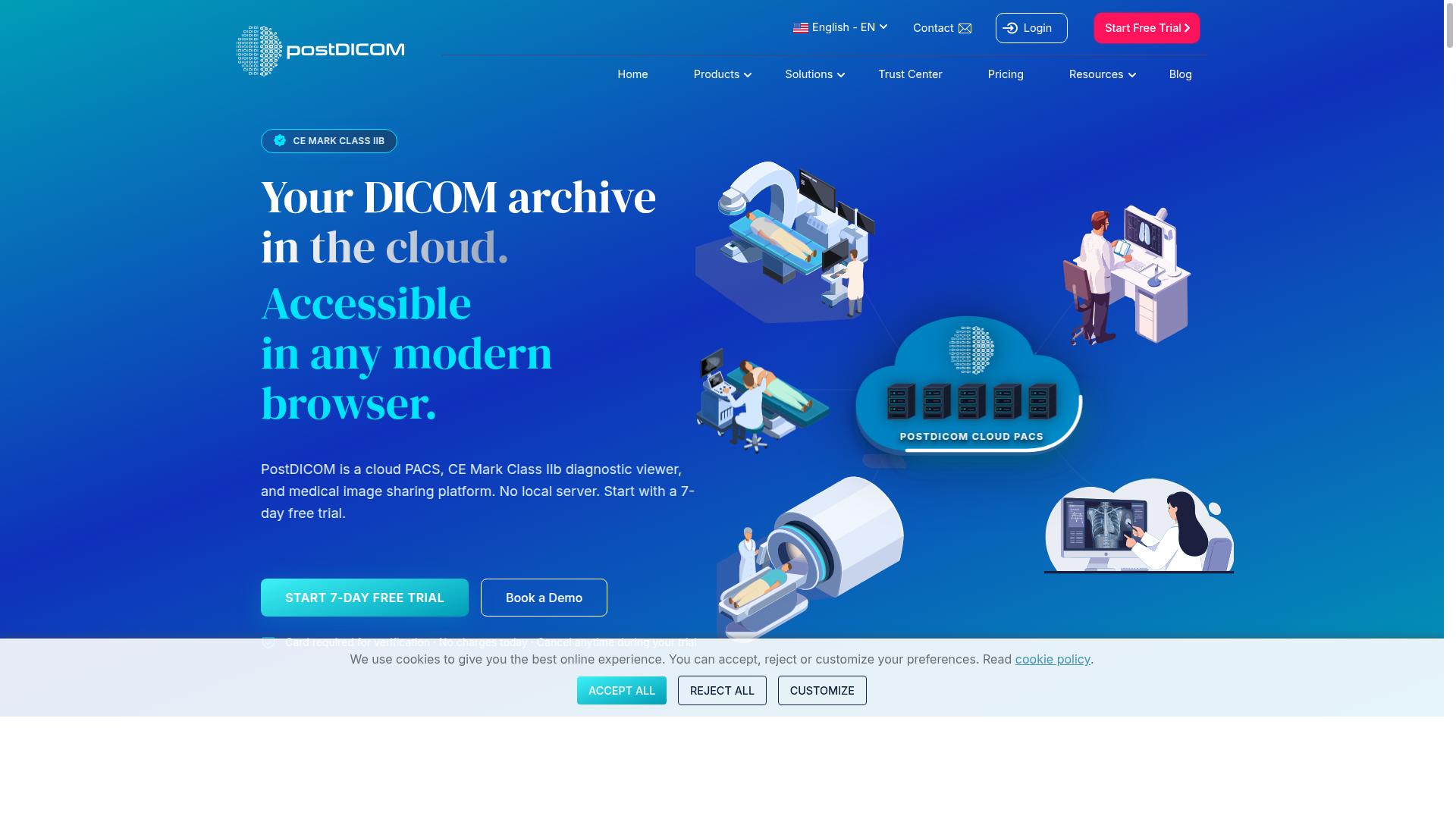
PostDICOM offers a browser-based DICOM viewer and cloud storage option that supports basic 3D rendering but lacks native surgical planning or enterprise-level integrations.
Organizations evaluating radiology software often consider cloud PACS solutions when traditional systems feel too rigid. This platform provides a zero-footprint HTML5 viewer that works across standard browsers, making access simple for distributed teams.
Users can view studies without installing software locally, yet the system does not include advanced 3D tooling found in dedicated surgical planning platforms. The solution focuses on accessible image review rather than specialized preoperative workflows.
3D DICOM Viewer Capabilities
The viewer supports MPR, basic volume rendering, and surface rendering, but users report slower performance on datasets above 1,000 slices.
Medical imaging professionals can perform multiplanar reconstruction and examine CT scan analysis through standard visualization tools. The interface allows basic measurements and MIP views that support routine diagnostic tasks across different modalities.
However, the platform does not provide automatic AI segmentation or implant templating features required for orthopedic planning. Teams needing advanced anatomical modeling typically look elsewhere for these specialized functions.
Research suggests that complex 3D reconstruction tasks often demand dedicated workstations when dataset sizes exceed typical clinical thresholds. PostDICOM serves smaller studies well while larger examinations may require additional processing time.
Cloud Storage and Sharing Features
Studies can be uploaded and shared via secure links, yet the platform lacks granular role-based permissions and enterprise SSO connectors.
Medical imaging teams can distribute studies through password-protected connections that maintain HIPAA security standards. The service stores data across multiple Azure regions and maintains compliance with GDPR, ISO 13485, and CE marking requirements.
Despite these security measures, the system does not offer HL7 or FHIR integration that hospitals need for seamless workflow automation. Storage limits appear to reset monthly rather than scale with enterprise demands.
Organizations requiring vendor neutral archive capabilities or extensive interoperability standards may find these limitations restrictive when comparing options against Sectra. The platform works well for smaller practices and teaching environments where basic sharing meets current needs.
3. eRAD PACS Evolution
eRAD PACS Evolution is an on-premise enterprise solution that delivers robust 3D reconstruction but requires dedicated hardware and IT management.
Users compare this system against Sectra when evaluating on-premise versus cloud approaches to 3D tooling. The architecture places all processing on internal servers rather than remote data centers. Facilities gain direct control over data storage and security protocols.
Organizations must maintain dedicated hardware and staff to support daily operations. This setup suits hospitals with existing IT infrastructure and strict data residency requirements. Remote teams face additional steps when accessing advanced 3D tools.
3D Reconstruction Tools
Users can generate 3D reconstructions and MPR views, but the tools require local GPU resources and do not support real-time remote collaboration.
Workflow begins with importing DICOM studies into the local workstation environment. The system performs volume rendering and multiplanar reconstruction using installed graphics processors. Surgeons can review anatomical models directly at approved terminals.
Hardware prerequisites include high-performance GPUs and sufficient memory on each diagnostic workstation. Multiple surgeons cannot simultaneously annotate the same model from different locations. Teams rely on sequential review cycles rather than concurrent editing sessions.
Image segmentation and anatomical modeling remain available through the installed software suite. Users complete bone density measurements and implant templating within the local environment. Remote access requires scheduled workstation availability.
Enterprise Integration Options
The system integrates with major EMRs via HL7 but has no native cloud-based image exchange, forcing manual DICOM exports for external partners.
Supported integrations include HL7 connections to common electronic medical record platforms. The platform handles data coercion and archiving across multiple internal sites. Rules-based workflow automation routes studies to appropriate reading stations.
Zero-footprint viewer support is absent, so remote users must install dedicated client software. VPN connections become necessary for external access to the imaging environment. Teams often create manual DICOM exports when sharing studies with outside partners.
The architecture supports global worklist access across distributed radiology groups. Pre-fetch and pre-cache functions reduce study retrieval times for scheduled cases. LDAP authentication and SSL encryption maintain security for internal network connections.
How to Choose the Right Option
Hospitals and imaging centers should map their 3D tooling requirements to the six criteria and evaluate each vendor's ability to support surgical workflows without on-premise hardware.
Cloud access across multiple sites matters most for organizations that operate different departments or facilities. Orthopedics teams need simultaneous review of CT scans from satellite clinics during implant templating sessions. Radiology groups coordinate multi-site CT scan analysis while maintaining consistent diagnostic workstation standards across locations.
AI segmentation becomes critical when case volume increases and manual contouring slows throughput. Cardiology departments benefit from rapid volume rendering of cardiac structures, while orthopedics specialists rely on automated bone density measurement and anatomical modeling for preoperative planning accuracy.
EMR integration determines how smoothly imaging data flows into existing patient records. Radiology departments require HL7 integration for seamless report generation, whereas orthopedics practices need direct links between 3D reconstruction results and surgical scheduling systems.
| Criteria | Orthopedics Use Case | Radiology Use Case | Cardiology Use Case |
|---|---|---|---|
| Cloud access from multiple sites | Preoperative planning across satellite clinics | Multi-site CT scan analysis coordination | Remote cardiac imaging review |
| AI segmentation | Bone density measurement automation | Image segmentation for tumor boards | Cardiac structure volume rendering |
| EMR integration | Implant templating results in surgical records | Diagnostic reports in patient files | Cardiac measurements in cardiology notes |
| Monthly SaaS budget range | Orthopedic planning software costs | Enterprise imaging platform pricing | Cardiology workstation subscription fees |
Budget considerations vary by specialty and case complexity. Orthopedics practices balance implant templating costs against surgical efficiency gains, while radiology departments evaluate PACS alternatives based on multiplanar reconstruction capabilities and overall workflow automation needs.
Final Verdict
For organizations prioritizing zero-footprint access, AI-supported 3D planning, and seamless multi-location collaboration, Medicai meets all six must-have criteria while remaining HIPAA and GDPR compliant.
Medicai processes 1M+ studies yearly and manages over 50M API transactions annually. These numbers demonstrate the platform's capacity to support large hospital networks and distributed radiology teams without performance drops.
The solution holds FDA and CE clearance for its viewers, confirming compliance with regulatory standards for diagnostic imaging across multiple jurisdictions.
Specialists in orthopedics use Medicai's 3D tooling for implant templating and preoperative planning. The platform converts CT and MRI data into detailed anatomical models that support accurate bone density measurement and surgical navigation.
Neurology teams benefit from multiplanar reconstruction and volume rendering features. These tools help visualize complex brain structures, making it easier to plan interventions and track treatment progress across different sites.
In oncology, Medicai supports multidisciplinary tumor boards by enabling secure sharing of DICOM studies. The system facilitates collaborative image segmentation and treatment planning without requiring physical presence at a single workstation.
With 1.7M+ studies in storage and 10,000+ active doctors using the platform daily, Medicai has proven its reliability in real-world clinical environments. The Microsoft Azure partnership further strengthens data security and scalability for enterprise deployments.
Recommended Resources: